¡Ciencia en las Fronteras!
Palabras clave:
microscopía, técnicas analíticas, microscopía electrónica, barrido, transmisiónResumen
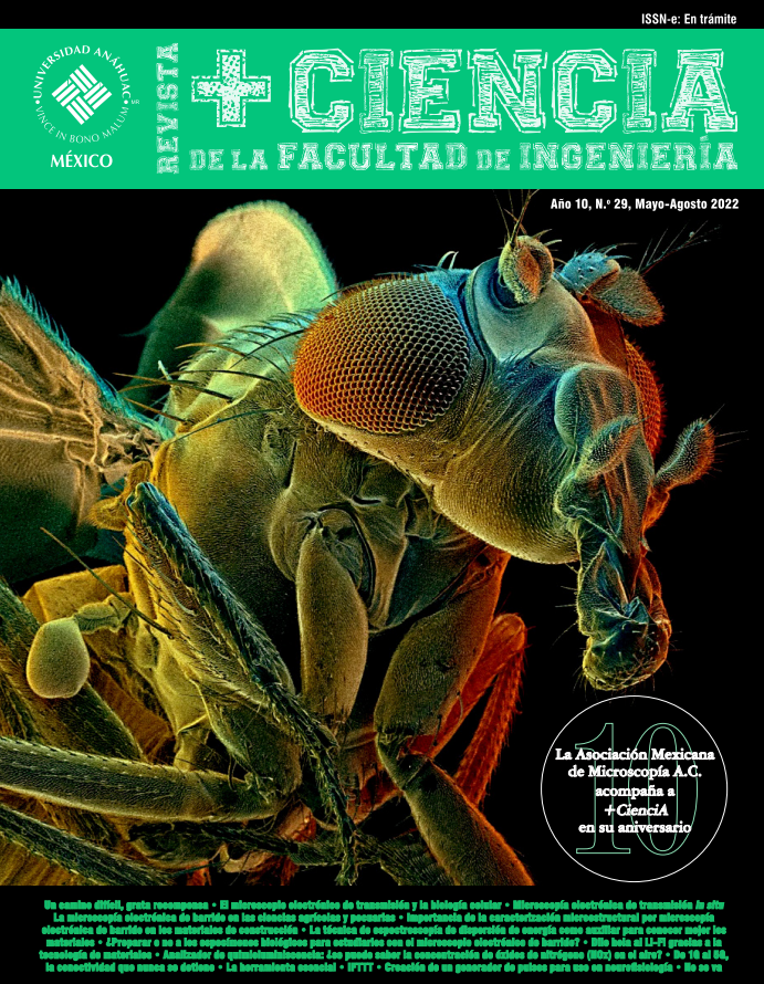
Edición especial de la sección en colaboració con la Asociación Mexicana de Microscopía A.C., (AMM).
Descargas
Referencias
Alberts, B., Johnson, A., Lewis, J., Morgan, D., Raff, M., Roberts, K., & Walter, P. (2015). Molecular biology of the cell, 6th ed. Garland Science.
Fawcett, D. W. (1981). The cell, 2nd ed. Saunders.
Marton, L. (1934). Electron microscopy of biological ob[1]jects. Nature, 133, 911.
Pollard, T. D., Earnshaw, W. C., Lippincott-Schwartz, J., & Johnson, G. T. (2017). Cell Biology, 3rd ed, Elsevier.
Porter, K. R., & Bonneville M. A. (1973). Fine structure of cells and tissues, 4th ed. Lea and Febiger.
Ochoa et al. (2019). Nanopart. Res., 21 (1).
Ortega et al. (2018). Microsc. Microanal, S1, 24, 952.
Ortega et al. (2018). AIP Advances 8. 056813.
https://nube.siap.gob.mx/gobmx_publicaciones_siap/pag/2021/Panorama-Agroalimentario-2021
Rivera-Jiménez, M., Zavaleta-Mancera, H., RebollarAlviter, A., Aguilar-Rincón, V., García-de-los-Santos, G., Vaquera-Huerta, H. et al. Phylogenetics and histology provide insight into damping-off infections of ‘Poblano’ pepper seedlings caused by Fusarium wilt in greenhouses. Mycological Progress (2018), 17(11):1237-1249.
Delgado-García, E., Cibrián-Tovar, J., González-Camacho, J., Valdez-Carrasco, J., Terán-Vargas, A., AzuaraDomínguez, A. Caracterización Morfológica de las Sensilas Antenales de Rhyssomatus nigerrimus (Coleoptera: Curculionidae). Southwestern Entomologist (2016), 41(1):225-240.
Gómez-Jaimes, R., Nieto-Ángel, D., Téliz-Ortiz, D., Mora-Aguilera, A., Martínez-Damián, M., Vargas-Hernández, M. Evaluación de la calidad e incidencia de hongos en frutos refrigerados de zapote mamey (Pouteria sapota (Jacq.) H. E. Moore and Stearn. Agrociencia (2009), 43(1):37-48.
Carrillo-Lopez, L., Huerta-Jimenez, M., Garcia-Galicia, I., Alarcon-Rojo, A. Bacterial control and structural and physicochemical modification of bovine Longissimus dorsi by ultrasound. Ultrasonics Sonochemistry (2019). 58: 104608.
González-Muñoz, S., Zavaleta-Mancera, H., MirandaRomero, L., Loera-Corral, O, Pinos-Rodríguez, J., Campos-Montiel, R. et al. Histochemical changes in early and mature Festulolium and maturation’s effects on rumen bacteria activity and in vitro degradation. Grassland Science (2018), 65(1): 23-31.
Lira‐Casas, R., Efren Ramirez‐Bribiesca, J., Zavaleta‐Mancera, H., Hidalgo‐Moreno, C., Cruz‐Monterrosa, R., Crosby‐Galvan, M. et al. Designing and evaluation of urea microcapsules in vitro to improve nitrogen slow release availability in rumen. Journal of the Science of Food and Agriculture (2018)
Khatib, J. (2016). Sustainability of construction materials. Second edition, Woodhead Publishing.
Zhou, W., et al. (2006). “Fundamentals of scanning electron microscopy (SEM)”. Scanning microscopy for nanotechnology. Springer, 1-40.
Bautista-León, F. (2017). Evaluación de la durabilidad de matrices de Cemento Portland, con adición de Mucílago de Nopal [Tesis de licenciatura]. Morelia, UMSNH.
Ramírez-Arellanes, S., et al. (2012). “Propiedades de durabilidad en concreto y análisis microestructural en pastas de cemento con adición de mucílago de nopal como aditivo natural”. Materiales de Construcción, 62(307): 327-341.
Zhou, W., Apkarian, R., Wang, Z. L. & Joy, D. (2007). “Fundamentals of scanning electron microscopy (SEM)”. Scanning Microscopy for Nanotechnology: Techniques and Applications. https://doi.org/10.1007/978-0-387-39620-0_1
Salman, A. (2020). Application of Nanomaterials in Environmental Improvement. Nanotechnol. Environ, 1-20. https://doi.org/10.5772/intechopen.91438
Schneider, R. (2011). “Energy-Dispersive X-Ray Spectroscopy (EDXS)”. Surface and Thin Film Analysis: A Compendium of Principles, Instrumentation, and Applications, second edition. https://doi.org/10.1002/9783527636921.ch18
Hodoroaba, V. D. Energy-dispersive X-ray spectroscopy (EDS). (2019). Characterization of Nanoparticles: Measurement Processes for Nanoparticles. Elsevier. https://doi.org/10.1016/B978-0-12-814182-3.00021-3
Bozzola, J. J. & Russell, L. D. (1999). Electron microscopy: Principles and techniques for biologists. 2nd ed. Jones and Bartlett Publisher. https://doi.org/10.22201/ceiich.24485691e.2020.25.69610
Douglas, B. (2001). Critical Point Drying of Biological Specimens for Scanning Electron Microscopy. Methods in Biotechnology, vol. 13, Supercritical Fluid Methods and Protocols. Edited by: JR Williams and AA Clifford. Humana Press Inc.
Echlin, P. (2009). Handbook of sample preparation for scanning electron microscopy and X-Ray microanalysis. Springer Science.
Hawkes, P. W. & Spence, J. C. (2007). Science of Microscopy, vol. 1. Springer Science. Reyes, G. J. Breve reseña histórica de la microscopía electrónica en México y en el mundo. Mundo Nano, 13 (25), 79-100.
Schatten, H., Pawley, J. B. (2008). Biological Low-Voltage Scanning Electron Microscopy. Springer Science.An introduction to electron microscopy. (Published on Jul 15, 2010). FEI Co.
Descargas
Publicado
Número
Sección
Licencia

Esta obra está bajo una licencia internacional Creative Commons Atribución-NoComercial-CompartirIgual 4.0.
+Ciencia por Universidad Anáhuac México se distribuye bajo licencia internacional Licencia Creative Commons Atribución-NoComercial-CompartirIgual 4.0 Internacional.


